By ZÁJER, P. SZENTMIKLÓSI, GY. SEBESTYÉN and G. KÖKÉNY
Department and Clinic of Surgery and Ophthalmology,
University of Veterinary Science Pharmaceutical
and Chemical Works CHINOIN Ltd.,
and Institute of Pharmacology, Semmelweis University Medical School, Budapest
(Received March 11, 1980)
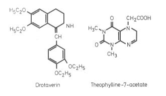
The first drugs used in medical practice for smooth muscle spasmolysis had been opium extracts. Of the opium alkaloids, papaverine was recognized as the most efficient spasmolytic. Later, MÉSZÁROS et al. (1961) developed the hydrated papaverine derivative Drotaverin, which proved to be superior even to papaverine, and was, after appropriate pharmacological testing, made available commercially under the trade name No-Spa® The spasmolytic, and coronary and peripheral vasodilator effects of Drotaverin have since been widely utilized in human clinical medicine. To prolong the duration of Drotaverin action, SZENTMIKLÓSI et al. (1977) developed Drotaverin-theophylline-7-acetate, which has become known under the trade~ name Depogen®. The wide use of spasmolytics in veterinary clinical medicine (colics in the horse, bronchial spasm, enteritis, etc.) prompted investigations into the effect of Depogen® on animals.
Experimental studies on the spasmolytic action of Drotaverin were performed by ISSEKUTZ (1963a, b) in rabbit intestinal loop, by HAUSSCHILD (1964) in rat and guinea pig intestine, and by DÁVID and GYARMATI (1962) in rat uterus. In these experiments, Drotaverin developed a more powerful spasmolytic action than papaverine.
We compared Drotaverin and Depogen® for duration of action by their influence on gastric emptying in conscious Beagle dogs.
Structural formula of Drotaverin-theophylline-7-acetate (trade name: Depogen).
| Group I | Control | not treated |
| Group II | Drotaverin pill | 20 mg/kg orally |
| Group III | Drotaverin injection | 10 mg/kg i.m. |
| Group IV | Depogen® substance | 30 mg/kg orally |
| Group V | Depogen® injection | 15 mg/kg i.m |
Zájer et al.
The above doses were established in preliminary experiments, under consideration of the molecular weights of the Drotaverin (419 daltons) and theophylline-7-acetate (238 daltons) components. Since Depogen® contains 64% Drotaverin and 36% theophylline-7-acetate,15 mg of it is equivalent to 10 mg Drotaverin. The oral doses of each drug were twice as large as the parenteral doses.
Ten treatments were performed in each group, in waking state. The dogs were deprived of feed and drinking water for the preceding 24 hrs. Twenty min after drug administration 20 ml/kg Novobarium (80 g Novobarium in 100 ml water) was given as contrast medium through a gastric tube. Thirty min and 1, 2, 4, and 6 hrs after administration of the contrast medium abdominal X-ray pictures were taken in the dorso-ventral view with a Contrastor 150 type apparatus (MEDICOR, Budapest), at 1 m focus distance, at 44 kV, 120 mA,
 |
 |
 |
 |
||
33 ms, on ORWO HS-11, 30 × 40 film. The roentgenographs were copied to photographic film and processed to 9 × 12 cm positive pictures.
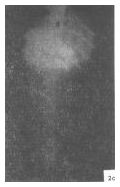
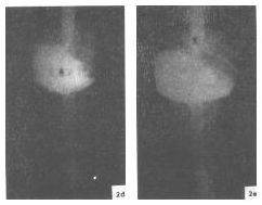
The effect of the drugs on gastric emptying was followed up by comparing the amount of contrast medium present in the stomach to a series of pictures prepared without drug administrations. The estimated difference was expressed in per cent. In preliminary rat experiments, administration of 2 ml contrast medium for 100 g body weight completely filled the stomach. Taking the quantity of contrast medium required for complete filling of rat stomach as 100%, 20, 40, 60, 80 and 100% gastric filling was produced in an anaesthetized in rising sequence by gradual administration of 2.0 ml/100 g (i.e. 5 × 0.4 m1/100 g) contrast medium. The radiographs taken after each administration were compared to reference pictures (Fig. 2a, b, c, d, e). Estimation of gastric filling in per cent is possible in a genetically homogeneous breed, where the shadow of the stomach shows practically no individual variation. The contrast medium having passed to the intestines was also taken into consideration in estimation, but the traces retained between gastric mucosal folds outside the homogeneous shadow were disregarded. Subjective error was greatly diminished by independent "blind" evaluation by 5 examiners, and disregard at final evaluation of estimates deviant from the most frequent one, as proposed by BARANSKI et al. (1964) for digestive function studies in astronautes; GÁTI et al. (1975) found the same approach satisfactorily conclusive in rats and we found it a suitable method for gastro intestinal motility studies in the same species.
In 3 rats of each of Groups II, III and IV, gastric emptying was also followed up by roentgenography 30 min after drug administration.
The percentual data of gastric emptying at different sampling intervals are shown in Table I. Five pictures for each of 50 treatments were evaluated.
Mathematical-statistical analysis of intergroup differences between drug treated and control series was carried out with the method of MANN and WHITNEY (cit. HAJTMAN, 1971) (Table II).
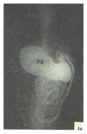
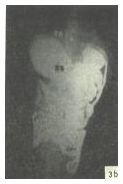
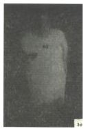
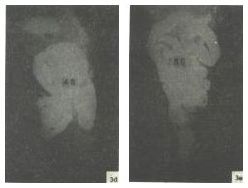
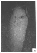
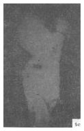
Control dogs not treated with drug (Group I) showed a gradual emptying within 4-6 hrs transit time; in no case was contrast medium demonstrable after 6 hrs (Figs 3a-e).
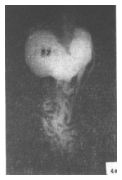
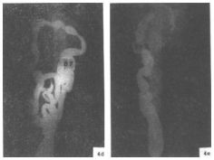
The, dogs treated orally (Group II) and parenterally (Group III) with Drotaverin showed a considerable increase in the rate of gastric emptying over the control, and a greater individual variation (Figs 4a-e).
Table I.
Estimated percentages of gastric filling in dogs treated and not treated with spasmolytics
| Groups | |||||
| Time after administration of contrast medium | |||||
| 0.5 | 1 | 2 | 4 | 6 | |
| hours | |||||
| I Control | 88 | 68 | 44 | 20 | 0 |
| II Drotaverin, oral | 82 | 42 | 38 | 14 | 10 |
| III Drotaverin i.m. | 70 | 38 | 18 | 4 | 2 |
| IV Depogen®, oral | 96 | 92 | 76 | 38 | 18 |
| V Depogen®, i.m. | 86 | 62 | 36 | 8 | 2 |
| Group | |||||||
| Level of gastric filling, per cent | |||||||
| 0 | 20 | 40 | 60 | 80 | 100 | ||
| 0.5 h | |||||||
| I | - | - | - | 1 | 4 | 5 | |
| II | - | - | - | 2 | 5 | 3 | |
| III | - | - | 3 | 1 | 4 | 2 | |
| IV | - | - | - | - | 2 | 8 | + |
| V | - | - | - | - | 7 | 3 | |
| 1 h | |||||||
| I | - | - | - | 6 | 4 | - | |
| II | - | - | 3 | 4 | 2 | 1 | |
| III | 1 | 3 | 4 | 1 | - | 1 | § 0 |
| IV | - | - | - | 1 | 2 | 7 | § 0 + |
| V | - | - | 1 | 7 | 2 | - | + x |
| 2 hrs | |||||||
| I | - | - | 8 | 2 | - | - | |
| II | - | 5 | 2 | 2 | 1 | - | |
| III | 5 | 3 | 1 | - | 1 | - | |
| IV | - | - | 1 | 2 | 5 | 2 | § 0 + |
| V | - | 1 | 9 | - | - | - | + |
| 4 hrs | |||||||
| I | 2 | 6 | 2 | - | - | - | |
| II | 6 | 2 | 1 | - | 1 | - | |
| III | 9 | - | 1 | - | - | - | |
| IV | 2 | 1 | 3 | 4 | - | - | + |
| V | 6 | 4 | - | - | - | - | § |
| 6 hrs | |||||||
| I | 10 | - | - | - | - | - | |
| II | 6 | 3 | 1 | - | - | - | |
| III | 9 | 1 | - | - | - | - | |
| IV | 5 | 1 | 4 | - | - | - | |
| V | 9 | 1 | - | - | - | - | |
 |
 |
 |
 |
||
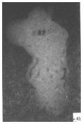
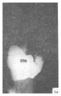
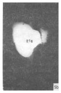
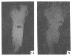
Dogs orally treated with Depogen® (Group IV) showed a considerable slowing-down of gastric emptying relative to control and Drotaverin-treated animals as well; generally, 50% of the contrast medium was still in the stomach at 6 hrs (Figs 5a-e).
Parenteral treatment with Depogen® (Group V) also accounted for a greater slowing-down of gastric emptying than, namely, parenteral, treatment with Drotaverin as shown by a greater retention of contrast medium at 4 and 6 hrs.
Follow-up of gastric emptying by roentgenography after oral and parenteral administration of Drotaverin showed that no emptying took place
 |
 |
 |
 |
||
in the first 20-25 min, but then its rate increased considerably over the control. Receptive relaxation was still shorter, about 10-15 min after parenteral administration of Depogen® (Group V), but no abrupt rise of emptying rate followed.
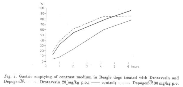
Oral administration of 20 mg/kg Drotaverin did not significantly alter the rate of gastric emptying relative to the control.
Ten mg/kg Drotaverin (Group III) administered i.m. within 1 h after the contrast medium gave rise to a significant acceleration of gastric emptying
 |
 |
 |
 |
||
relative to control and oral Drotaverin treatment, owing to vegetative counter-regulation in response to termination of motility inhibition (receptive relaxation) after 20-25 min. X-ray examination detected acceleration of gastric emptying after oral administration Drotaverin as well.
Oral administration of 30 mg/kg Depogen® (Group IV) accounted for a significant slowing-down of gastric emptying relative to parenteral Drotaverin within 30 min, relative to control and oral and parenteral Drotaverin within 1-2 hrs, and relative to parenteral Drotaverin and parenteral Depogen® within 4 hrs, after administration of the constrast medium. This indicated a lasting inhibitory action of the oral Depogen® preparation on smooth muscle motility.
Parenteral treatment with 15 mg/kg Depogen® (Group V) caused a significant slowing-down of gastric emptying for 2 hrs after contrast medium only in relation to parenteral Drotaverin (Group III) which alone accelerated the rate of emptying over normal. No abrupt increase of gastric emptying followed upon the initial 10-15 min period of receptive relaxation, owing presumably to prevention of increased counter-regulation by long-term Drotaverin action.
In conclusion, oral and i.m. treatment of conscious dogs with 10 and 20 mg/kg Drotaverin, respectively, inhibited gastric emptying for 20-25 min where after emptying proceeded at an increased rate, owing to acceleration by vegetative counter-regulation. Orally administered 30 mg/kg Depogen® prolonged the inhibition of gastric emptying significantly, for more than 2 hrs, relative to the control and to Drotaverin group as well.
Inhibition of gastric motility by Depogen® was not followed by an abrupt increase in the rate of emptying, indicating that this drug acted longer than Drotaverin.
Further to enabling the comparative evaluation of two preparations for duration of action, the present studies have substantiated the suitability of the X-ray technique for gastro intestinal motility studies, and for the quantitative estimation of gastric emptying as well. Apart from common laboratory animals, genetically-even Beagle dogs may be used in such studies. Since the X-ray technique does not interfere with physiological gastro intestinal passage, it should be, as proposed earlier by KOVÁCH (1965), preferred to the balloon technique whenever possible.
Beagle dogs starved for 24 hrs were treated with Drotaverin (No-Spa®) and Depogen®, orally at 20 and 30 mg/kg, i.m. at 10 and 15 mg/kg dose level, respectively, in groups of 10, with one group used as untreated control. Twenty min after drug treatment, 20 ml/kg Novobarium was administered as contrast medium through a gastric tube and 0.5, 1, 2, 4 and 6 hrs later roentgenograms were taken of the abdominal cavity to estimate the influence of the drugs on gastric emptying by comparison to a reference series of radiographs taken at 20 to 100% levels of gastric filling. The estimated percentual differences between gastric emptying with and without spasmolytic treatment were analyzed for statistical significance by the method of MANN and WHITHNEY.
Drotaverin inhibited gastric emptying for 20-25 min, upon which followed a considerable increase of emptying rate over the control. Reactive acceleration failed to take place after relaxation induced by parenteral administration of Depogen®, and oral Depogen® treatment slowed down gastric emptying relative to the control and the longer duration of the Dpogen's action.
Address of the senior author: Dr. József ZÁJER, 1078, Budapest, Landler J. u. 2., Hungary.